Skin check for skin cancer.
Skin cancer diagnosis is now relatively straight forward. Thank you very much to the Skin cancer college of Australia, University of QLD, Sydney university and Drs like Dr Peter Bourne, Dr Scott Menzie, Dr Dr
More cancer are missed by not looking.
We know quite a number of people who have died because of melanoma. Most of them would still be alive if their melanoma was not missed. So if you see an ugly duckling mole on your skin – see you doctor immediately.
Skin cancer examination is so quick and simple. We use big broad bright light and magnification and selected microscopy to examine your skin. Skin cancer examination takes less than 3 minutes ! May be, 10 minutes if the doctor is still in training and, a lot lot more if the doctor biopsy every anxious lesions. (extra time is needed for undressing & re-dressing )
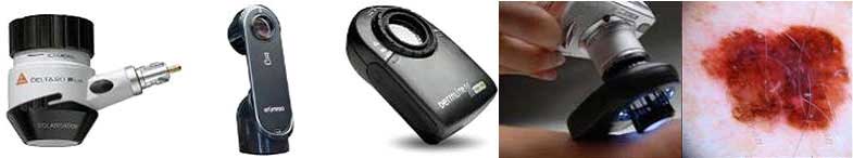
At our Crown kin cancer clinic, we use our clinical experience together with dermoscopy to do skin checks.
Dermoscopy is a simple method that enables detailed examination of the structure of skin lesions or moles. It improves the diagnosis of the vast majority of all skin cancer types without resorting to excessive biopsies– saving you time, unnecessary stress and costs.
Thanks to Aussies skin cancer doctors, the like of Dr Scott Menzie, Dr Dr
Micro-photo’s of moles can be taken through these devices for later review if indicated.
There are many eye catching and elaborate skin check or mole check machines available commercially. They are good for the inexperienced . They are not 100% accurate . The ideal person to operate mole photo machine are ones who have good working knowledge of dermoscopy. Ultimately, clinicians serve as the final arbiters of the diagnosis for examined lesions.
Photographic documentation may be of value if you are at high risk for developing melanoma, i.e. if you have multiple moles and numerous dysplastic neavii ( atypical moles ) where biopsies of them all would be impractical and hence photographic follow up would be better.

Basal cell carcinoma. This is a glaring example of a bcc. The clinico-dermoscopic picture is so obvious that biopsy is unnecessary. On the left is the naked eye image of a bcc and it’s microscopic picture on the right.

Melanoma. Both of these lesions look suspicious. The novice would biopsy both. The expert dermoscopist would biopsy the left lesion based on the clinical history and the dermoscopic image. The right lesion is a benign clotted heamangioma- “blood blister”. The lesion on the left is a early stage melanoma.

This patient was told that this lesion was a squamous cell carcinoma, A novice would incline to believe it. On history taking, general skin check and dermoscopic examination, a diagnosis of varicose eczema was made. We treated his varicose veins by ultrasound guided sclerotherapy and the rash and ulcer resolved.